


Demystifying AVM: Symptoms, Diagnosis, and What to Expect
Reviewed by: Manipal Hospitals
Posted on
7 Min Read

Not sure what you’re looking for?
Get in touch with our representatives for help!
Contact UsAn arteriovenous malformation (AVM) is an abnormal tangle of blood vessels where arteries connect directly to veins, bypassing the capillaries. Capillaries normally slow down blood flow, allowing oxygen and nutrients to be delivered to tissues. In an AVM, this crucial network is missing, leading to high-pressure blood flowing from arteries directly into weaker veins. This can cause the veins to enlarge and potentially rupture, leading to bleeding (haemorrhage) in the brain or other parts of the body.
Where Do AVMs Occur?
AVMs can develop anywhere in the body, but are most found in:
-
Brain
-
Spinal cord
-
Face and scalp
-
Arms and legs
-
Lungs
-
Liver and other internal organs
Brain AVMs are particularly important because they can cause bleeding within the brain.
What Causes AVMs?
The exact cause of an arteriovenous malformation is not fully understood, but most AVMs are believed to be congenital, meaning they are present at birth. They are generally not hereditary, and it is rare for them to be passed down through families. They are thought to arise from an error in foetal development, where the normal formation of blood vessels goes awry. While AVMs are present from birth, symptoms may not appear until later in life, sometimes even decades later, as the malformation grows or complications arise. Acquired AVMs are extremely rare and are usually a result of trauma or infection.
Recognising Arteriovenous Malformation Symptoms
The symptoms of an arteriovenous malformation vary widely depending on its size, location, and whether it has bled. Some individuals with AVMs may never experience symptoms, while others may have severe, life-altering complications. Knowing the symptoms of arteriovenous malformation helps with timely intervention.
Brain AVMs
Patients may experience:
-
Severe headaches
-
Seizures
-
Weakness of an arm or leg
-
Difficulty speaking
-
Visual disturbances
-
Sudden brain haemorrhage (stroke)
Peripheral AVMs (Limbs, Face, Scalp)
Symptoms may include:
-
Swelling
-
Pain
-
Warmth over the affected area
-
Visible pulsations
-
Skin discolouration
-
Bleeding from the lesion
-
Cosmetic deformity
Other AVM Locations
AVMs can also occur in other parts of the body, leading to location-specific symptoms:
-
Pulmonary AVMs (Lungs): Shortness of breath, fatigue, nosebleeds, and reduced oxygen levels.
-
Gastrointestinal AVMs: Chronic bleeding, leading to anemia, fatigue, and blood in stool.
-
Peripheral AVMs (Limbs): Swelling, pain, visible pulsation, and skin discoloration in the affected area.
Diagnosing Arteriovenous Malformations
Early and accurate diagnosis of an arteriovenous malformation helps prevent complications and plan effective arteriovenous malformation treatment. Due to the diverse nature of symptoms, diagnosis often begins with a thorough neurological examination and detailed medical history.
Diagnostic Tools and Procedures
Several advanced imaging techniques are used to accurately identify and characterise AVMs:
-
Magnetic Resonance Imaging (MRI): Via detailed images, MRI helps visualise the AVM's structure and size.
-
Magnetic Resonance Angiography (MRA): A specialised MRI that focuses on blood vessels, revealing the abnormal flow within the AVM.
-
Ultrasound with Doppler: It works by using sound waves to visualise blood vessels (B-mode) while simultaneously measuring the speed, direction, and volume of blood flow (Doppler).
-
Computed Tomography (CT) Scan: Can quickly detect acute bleeding and provide initial structural information.
-
Computed Tomography Angiography (CTA): A CT scan combined with an injected contrast dye to visualise blood vessels and blood flow patterns.
-
Cerebral Angiography (Digital Subtraction Angiography - DSA): In this procedure, a catheter is inserted into an artery (usually in the groin) and guided to the affected organ where contrast dye is injected to provide real-time, highly detailed X-ray images of the blood vessels. This allows experts to map the AVM's feeders, nidus (tangle), and draining veins precisely. This advanced angiography ensures precise arteriovenous malformation treatment planning.
Arteriovenous Malformation Treatment Options
Not every AVM requires immediate intervention. Treatment decisions depend on the location, size, symptoms, risk of bleeding and age and overall health of the patient. The goal is to prevent complications while preserving normal tissue function. Many AVMs are evaluated and managed through Neurological Vascular Intervention procedures, which focus on treating complex vascular abnormalities affecting the brain and nervous system. Some of the options include:
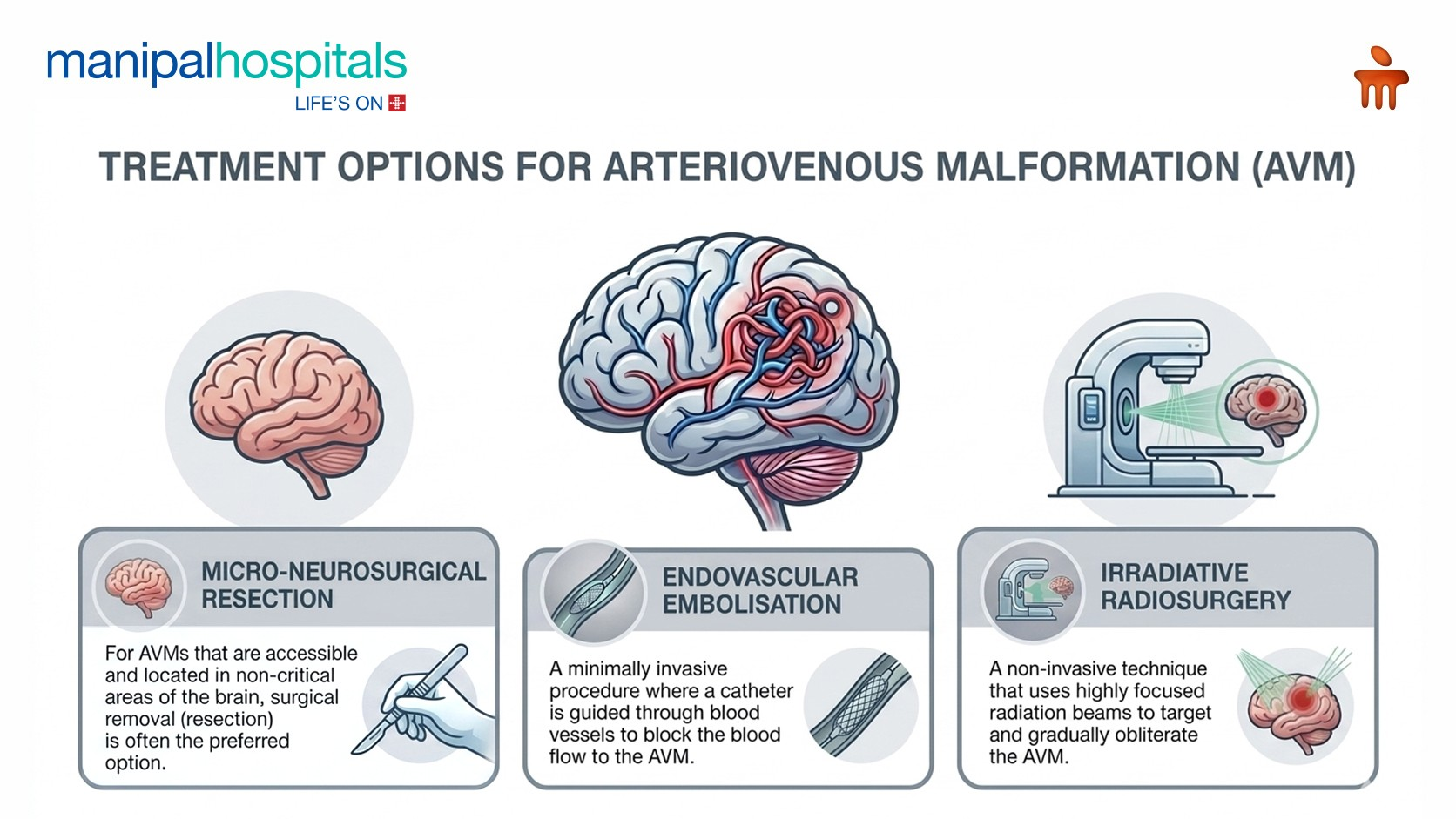
Micro-Neurosurgical Resection
For AVMs that are accessible and located in non-critical areas of the brain, surgical removal (resection) is often the preferred option. This procedure involves opening the skull (craniotomy) to directly visualise and surgically remove the AVM. The goal is complete removal to eliminate the risk of future bleeding.
Endovascular Embolisation
Endovascular embolisation is a minimally invasive procedure performed by an Interventional Radiologist where a catheter is guided through blood vessels to the AVM. A liquid embolic agent (like a medical glue) is then injected into the AVM to block blood flow. This procedure can be used as a standalone arteriovenous malformation treatment, particularly for smaller AVMs, or more commonly, as a pre-surgical step to reduce the AVM's size and blood flow.
Irradiative Radiosurgery
Stereotactic radiosurgery (SRS) is a non-invasive technique that uses highly focused radiation beams to target and gradually obliterate the AVM. It is often used for smaller AVMs (typically less than 3 cm), deeply located, or in critical areas where traditional surgery is too risky.
Combined Approaches and Observation
Sometimes, a combination of these treatments may be used to achieve the best outcome. For example, embolisation might be followed by surgical resection or radiosurgery. In some cases, particularly for very small, asymptomatic AVMs with a low risk of rupture, a "watch-and-wait" approach with regular monitoring might be recommended.
The use of Interventional Radiology has revolutionised AVM management. Many lesions that once required major surgery can now be treated through tiny catheters under image guidance. This approach offers patients, lesser pain, shorter hospital stay, faster return to daily activities, and improved outcomes.
When Should You Seek Medical Attention?
Consult a specialist if you experience:
-
Unexplained swelling or pulsatile masses
-
Recurrent bleeding from a vascular lesion
-
Persistent headaches with neurological symptoms
-
Seizures without an obvious cause
-
Sudden weakness, speech difficulty, or vision loss
Conclusion
Arteriovenous Malformations are uncommon but potentially serious vascular abnormalities. Advances in imaging and minimally invasive endovascular therapies have dramatically improved outcomes. With timely diagnosis and appropriate treatment, most patients can lead normal and productive lives.
If you or a loved one has been diagnosed with an AVM, consultation with a multidisciplinary team, including Interventional Radiology, Neurosurgery, and Vascular Specialists, at Manipal Hospital, Goa, can help determine the most effective treatment strategy.
Frequently Asked Questions
The most dangerous complication of an AVM is haemorrhage, which can lead to stroke, severe neurological damage, or even be life-threatening.
Most AVMs are congenital (present at birth) but are generally not hereditary, meaning they are rarely passed down through families.
Yes, AVMs can slowly grow larger over time. This growth can increase the risk of rupture and the severity of symptoms.
Recovery time varies greatly depending on the type of treatment, the AVM's location, and whether there were complications. It can range from a few weeks for less invasive procedures to several months or even longer with rehabilitation after a haemorrhage or complex surgery.
After complete surgical resection or successful radiosurgical obliteration, the recurrence of an AVM is rare. However, follow-up imaging is essential to confirm complete eradication and monitor for any new developments.
Share this Article


Recent Blogs

Postpartum Haemorrhage: Causes, Symptoms & Emergency Signs
29 Jun 20267 Min Read
Delivering a baby is a major physical event that puts a lot of demand on a woman's body. While the focus is often on the arrival of the baby, the management of the third stage of labour—delivering the placenta and controlling blood loss—is just as crucial to a safe delivery. Understanding the risks during this window, including excessive bleeding or postpartum haemorrhage, is a vital part of maternal care.
Read NowDislocated Elbow: Signs, First Aid & Recovery Time
26 Jun 20267 Min Read
An elbow dislocation can be a frightening and painful experience, often occurring unexpectedly due to falls or sports injuries. While it's a serious injury, understanding the signs, knowing the correct first aid, and following a structured recovery plan can significantly improve outcomes.
Read NowUnderstanding Chronic Diarrhoea: More Than Just a Temporary Stomach Upset
26 Jun 20266 Min Read
Chronic diarrhoea, characterised by loose, watery stools occurring three or more times a day for a period exceeding four weeks, is a common and often debilitating condition affecting millions worldwide. While acute diarrhoea is usually short-lived and often caused by infections, chronic diarrhoea points towards a more persistent underlying issue that requires careful investigation.
Read NowSoothing the Itch: Expert Solutions for Itchy Inner Thighs
22 Jun 20267 Min Read
Experiencing persistent itching in your inner thighs can be incredibly uncomfortable, disruptive, and even embarrassing. While often a common annoyance, it can sometimes signal an underlying skin condition that requires attention. Understanding the common causes of itchy inner thighs will help explore effective remedies and adapt practical prevention tips to keep your skin healthy and comfortable.
Read NowCan an ECG Detect a Heart Attack? What the Test Really Shows
15 Jun 20268 Min Read
In moments of chest pain or suspected cardiac distress, an Electrocardiogram (ECG or EKG) is often the first diagnostic test performed. It's a quick, non-invasive procedure that provides a snapshot of your heart's electrical activity. But can an ECG detect a heart attack definitively? While an ECG is an indispensable tool in emergency cardiac care, its ability to pinpoint a heart attack is nuanced.
Read NowDemystifying AVM: Symptoms, Diagnosis, and What to Expect
12 Jun 20267 Min Read
An arteriovenous malformation (AVM) is an abnormal tangle of blood vessels where arteries connect directly to veins, bypassing the capillaries. Capillaries normally slow down blood flow, allowing oxygen and nutrients to be delivered to tissues. In an AVM, this crucial network is missing, leading to high-pressure blood flowing from arteries directly into weaker veins. This can cause the veins to enlarge and potentially rupture, leading to bleeding (haemorrhage) in the brain or other parts of the body.
Read NowKnee Pain When Bending: Causes, Symptoms, and Treatment Options
12 Jun 20266 Min Read
Knee pain is a common ailment, but when it specifically flares up while bending, it can be particularly debilitating, affecting daily activities from walking to climbing stairs. The knee is a complex joint, crucial for movement and supporting body weight. When you experience knee pain when bending, it signals an issue within its bones, ligaments, tendons, and cartilage. This discomfort can range from a dull ache to sharp, intense pain, often accompanied by other symptoms.
Read NowWhat are Bilateral Polycystic Ovaries?
05 Jun 202610 Min Read
The term bilateral polycystic ovaries often raises concerns and questions for many women. It refers to the presence of multiple small cysts on both ovaries, a characteristic feature often associated with Polyendocrine Metabolic Ovarian Syndrome (PMOS). While the name can sound alarming, understanding what bilateral polycystic ovaries are and its implications is the first step towards effective management. While it is a condition that requires attention and management, it is generally not life-threatening and many women lead full, healthy lives with proper care.
Read NowWhy Do I Feel Weak All the Time? Causes Explained
27 May 20267 Min Read
There are times when the “I am just tired” becomes “why do I feel weak all the time?” This persistent feeling of low energy, muscle fatigue, or an overall lack of strength can significantly affect daily life, making even routine tasks feel Herculean. While often used interchangeably, "weakness" and "fatigue" are distinct. Fatigue is a feeling of tiredness or lack of energy, whereas weakness (or asthenia) is a specific reduction in physical strength. When you experience true weakness, it means you can't exert the same force as before, or your muscles tire out much faster. This isn't just about feeling sleepy; it's about a noticeable decrease in your physical capacity. Studies find that a major portion of the adult population experiences unexplained persistent weakness at some point, highlighting the common nature of this weakness and the importance of seeking professional guidance. Persistent weakness should be evaluated by specialists in the Department of Internal Medicine to identify underlying health conditions and provide appropriate treatment.
Read NowInflammatory Breast Cancer: Early Signs You Shouldn’t Ignore
27 May 20266 Min Read
While many associate breast cancer with a palpable lump, some forms present differently. One such form is inflammatory breast cancer, a rare yet aggressive type that often doesn't start with a lump. Because its signs can be subtle and mimic common skin conditions, it's frequently misunderstood, leading to delayed diagnosis. Despite its rarity, the rapid progression of inflammatory breast cancer makes early detection vital. Understanding its unique presentation is your first line of defence. With timely intervention, significant strides have been made in improving outcomes. Specialists in the Department of Oncology play a crucial role in the early diagnosis, evaluation, and personalised treatment of inflammatory breast cancer.
Read Now