

Baner

4.3
Advanced imaging that combines metabolic and structural detail for precise diagnosis. You're in safe hands.
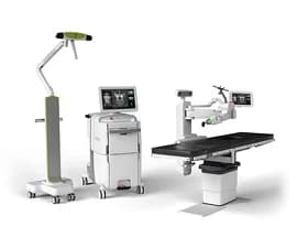
A PET-CT scan is an advanced imaging technology that combines Positron Emission Tomography (PET) and Computed Tomography (CT) in a single scan. It captures both the metabolic activity of cells and detailed structural images of organs and tissues, helping doctors detect disease earlier and more accurately. This PET-CT technology is widely used in cancer care for diagnosis, staging, treatment planning, and monitoring response to therapy, and is also valuable in cardiology and neurology. At Manipal Hospitals, the Nuclear Medicine department operates PET-CT services with specialist radiologists and nuclear medicine physicians, supporting faster, more informed treatment decisions across specialities.
Before the scan, a small amount of a radioactive tracer (usually a form of glucose) is injected into the patient's bloodstream. Since cancer cells and other abnormal tissues often use more glucose than healthy cells, they absorb more of the tracer.
The PET-CT scan machine then captures two sets of images in one sitting: the PET component detects areas of high tracer uptake (indicating abnormal activity), while the CT component provides a detailed anatomical map. These two images are combined to precisely pinpoint the location, size, and nature of any abnormality, helping doctors distinguish between benign and malignant conditions with far greater clarity than either scan alone.
4.3
The doctor reviews the patient's medical history and explains why a PET-CT scan is being recommended, along with what to expect.
| Advanced Technology | Conventional Approach | |
|---|---|---|
| Precision | High-precision hybrid imaging combines metabolic activity with detailed anatomical localisation. | Primarily focuses on structural size and shape, which may not detect early cellular changes. |
| Visualisation | 3D fusion imaging maps tracer uptake and metabolic activity directly onto high-resolution anatomical images. | Relies on separate structural images, offering limited insight into functional or metabolic activity. |
| Invasiveness | Non-invasive diagnostic imaging requiring only a standard intravenous radiotracer injection. | Non-invasive imaging may be followed by an invasive biopsy when further assessment is required. |
| Recovery | No downtime; patients can generally resume their normal activities after the scan. | No downtime from imaging, although additional scans or procedures may be needed when findings are inconclusive. |
| Patient Experience | A streamlined imaging process provides structural and metabolic information in a single session, helping reduce the need for multiple visits. | Multiple imaging appointments may be required to obtain both structural and functional information. |


Discover stories of
We leave no stone unturned in exploring alternate treatment options for our patients. Our multidisciplinary teams deliver advanced, personalised care focused on restoring health and empowering patients to overcome serious medical challenges and regain their lives.
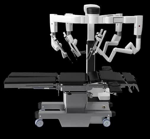
Robot Assisted Radical Prostatectomy
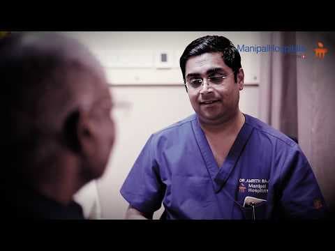
Robot Assisted Radical Prostatectomy
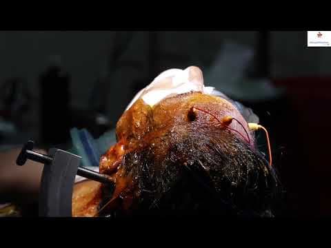
A miraculous Procedure, Robot Assisted Radical Prostatectomy done by Dr. Amrith Raj Rao, Consultant - Urology And Robotic Surgery...
With a legacy of several decades, Manipal Hospitals is one of India's leading multi-specialty healthcare networks. We deliver comprehensive outpatient, inpatient, emergency, and advanced surgical care tailored for patients of all age groups.

Not sure what you're looking for?
A PET-CT scan is a combined imaging test that merges PET and CT technology to show both the metabolic activity and structural detail of tissues in a single scan, helping doctors detect and evaluate disease with greater precision.
While a CT scan shows only the structure of organs and tissues, PET-CT technology adds a metabolic layer, highlighting areas of abnormal cell activity and giving doctors a more complete picture, especially for cancer diagnosis and staging.
Yes. The radioactive tracer used is given in a very small, carefully controlled dose, and the procedure is considered safe for most patients. Your doctor will review your specific health history before the scan.
The scan itself typically takes 20–40 minutes, though patients should plan for 2–3 hours total to allow time for tracer injection and uptake before the pet ct scan machine begins imaging.
Yes. Patients are usually asked to fast for several hours before the scan and avoid strenuous exercise the day before, as this can affect tracer uptake. Your care team will provide specific instructions.