

Baner

4.3
Early, accurate breast cancer screening for every stage of life. You're in safe hands.
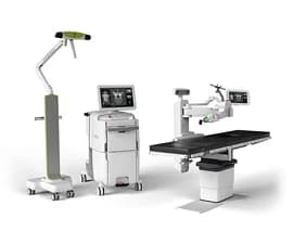
Mammography is a specialised imaging technique that uses low-dose X-rays to examine breast tissue, helping detect abnormalities such as lumps, calcifications, or early-stage tumours often before they can be felt. It is one of the most effective tools for early breast cancer detection, significantly improving treatment outcomes. At Manipal Hospitals, mammography is performed by the Radiodiagnosis department using a digital mammography machine, offering sharper imaging and lower radiation exposure than older analog systems. It supports both routine screening for women's health and diagnostic evaluation for patients with symptoms, working closely with the Breast Oncology team when further treatment is needed.
During the procedure, the breast is gently compressed between two plates on the digital mammography machine to spread the tissue evenly, allowing clearer, more detailed images with a lower radiation dose. Unlike traditional film mammography, digital images are captured electronically, allowing radiologists to zoom in, adjust contrast, and closely examine areas of concern on screen.
The entire scan typically takes just a few minutes per breast. Images are reviewed by a radiologist, who looks for lumps, unusual masses, or tiny calcium deposits (calcifications) that could indicate early-stage changes. If anything unusual is found, additional imaging or a biopsy may be recommended for further evaluation.
13+
Years of Experience
13+
Operational Hospitals
4.3
The doctor reviews the patient's age, family history, and any symptoms to determine if mammography is recommended for screening or diagnosis.
| Advanced Technology | Conventional Approach | |
|---|---|---|
| Precision | Captures multiple low-dose X-ray images from different angles to reconstruct a 3D volumetric view of dense breast tissue | Provides a flat, single 2D projection image, where overlapping dense tissue can obscure underlying lesions |
| Visualization | Multi-planar slice-by-slice rendering that isolates small, early-stage calcifications and subtle architectural distortions | Limited multi-angle visibility with a higher potential for tissue overlap artefacts in dense breasts |
| Invasiveness | Non-invasive diagnostic imaging with optimised radiation dosing protocols compliant with international radiation safety standards | Non-invasive, but may require additional follow-up views or supplementary imaging to resolve unclear areas |
| Recovery | Zero downtime; patients resume normal daily activities immediately following the procedure | Zero downtime; may require repeat exposure sessions if initial structural clarity is insufficient |
| Patient Experience | Faster acquisition times with ergonomic, curved compression paddles that automatically optimise pressure for improved comfort | Standard flat paddles requiring higher sustained compression pressure during longer scan hold times |


Discover stories of
We leave no stone unturned in exploring alternate treatment options for our patients. Our multidisciplinary teams deliver advanced, personalised care focused on restoring health and empowering patients to overcome serious medical challenges and regain their lives.
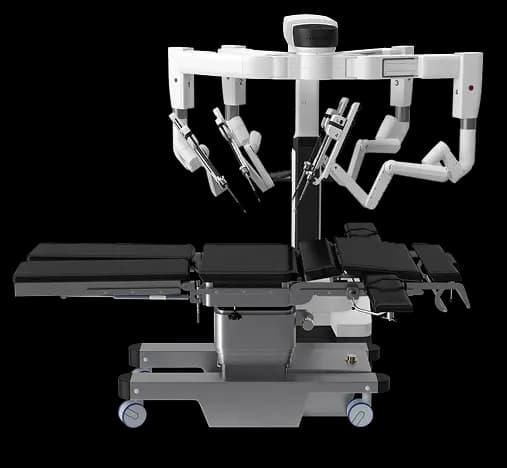
Robot Assisted Radical Prostatectomy
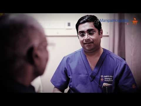
Robot Assisted Radical Prostatectomy
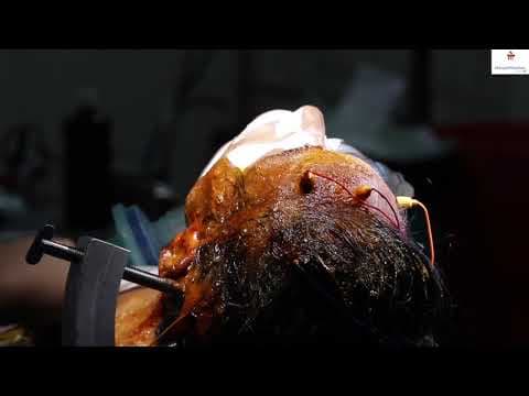
A miraculous Procedure, Robot Assisted Radical Prostatectomy done by Dr. Amrith Raj Rao, Consultant - Urology And Robotic Surgery...
With a legacy of several decades, Manipal Hospitals is one of India's leading multi-speciality healthcare networks. We deliver comprehensive outpatient, inpatient, emergency, and advanced surgical care tailored for patients of all age groups.

Not sure what you're looking for?
Mammography is used to screen for and detect breast abnormalities, including early signs of breast cancer, often before they can be felt during a physical exam.
Digital mammography captures breast X-ray images electronically instead of on film, allowing radiologists to view, zoom in on, and adjust images for clearer, more detailed analysis compared to traditional mammography.
Screening age recommendations vary, but many doctors suggest women begin routine mammography around age 40, or earlier if there is a family history of breast cancer. Your doctor can advise on the right schedule for you.
Some women experience brief discomfort or pressure during the breast compression, but it typically lasts only a few seconds and is well tolerated by most patients.
This depends on individual risk factors and doctor recommendations, but routine screening is often advised annually or every one to two years for women in the recommended age group.