

Baner

4.3
Precise Functional Imaging for Whole-Body Diagnosis. You're in safe hands!
A gamma camera is a specialised imaging device used in nuclear medicine to capture images of how organs and tissues are functioning. After a patient receives a small, safe dose of a radioactive tracer, the gamma camera radiology equipment detects the gamma rays emitted from within the body and converts them into detailed images that reveal areas of abnormal activity. This gamma ray imaging approach is especially valuable for detecting conditions that may not yet show up on a structural scan like a CT or MRI, including certain bone, thyroid, kidney, and cardiac conditions. At Manipal Hospitals, gamma camera imaging is performed by the Nuclear Medicine department and supports diagnosis across cardiology, oncology, endocrinology, and nephrology.
Before the scan, the patient is given a small amount of a radioactive tracer, usually through an injection, which travels through the bloodstream and accumulates in specific organs or tissues depending on the type of study being done. The gamma camera then detects the gamma rays released by this tracer as it decays inside the body.
Using a series of detectors, the gamma camera radiology system converts these emissions into a detailed image that shows exactly where the tracer has concentrated, highlighting areas of increased or decreased activity that may indicate disease. Unlike X-rays or CT scans, which show anatomy, gamma ray imaging shows function, making it possible to detect problems at a cellular or metabolic level, often before structural changes become visible on other scans.
The scan is painless and non-invasive, though it may take anywhere from 20 minutes to a few hours depending on the study, as time is needed for the tracer to circulate and concentrate in the target area.
1+
Years of Experience
1+
Operational Hospitals
4.3
The doctor reviews your symptoms and medical history and determines whether a gamma camera scan is needed to assess how a specific organ or system is functioning.
| Advanced Technology | Conventional Approach | |
|---|---|---|
| Precision | Captures 3D tomographic reconstructions with high spatial resolution to localise metabolic activity and deep-tissue functional anomalies | Limited to 2D planar projections, where overlapping physiological structures can obscure smaller or deeper functional lesions |
| Visualization | Multi-planar 3D slice rendering (SPECT) with cross-sectional mapping that maps radiotracer distribution in specific organ tissues | Single-angle static planar views with limited volumetric depth and reduced clarity in dense anatomical areas |
| Invasiveness | Non-invasive imaging performed via low-dose radiotracer administration compliant with strict nuclear medicine safety protocols | Non-invasive diagnostic process, but may require higher tracer doses or supplementary scans to resolve unclear planar views |
| Recovery | Zero downtime; radiotracers decay naturally and clear from the body quickly with standard post-procedure hydration | Zero downtime; patients may need extended waiting periods between repeated static acquisition runs |
| Patient Experience | Faster acquisition times using dual-head detectors that capture multiple angles simultaneously, reducing scan duration and motion artifacts | Longer acquisition times with single-detector rotation, requiring prolonged immobility on the imaging bed |


Discover stories of
We leave no stone unturned in exploring alternate treatment options for our patients. Our multidisciplinary teams deliver advanced, personalised care focused on restoring health and empowering patients to overcome serious medical challenges and regain their lives.
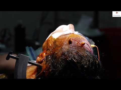
Robot Assisted Radical Prostatectomy
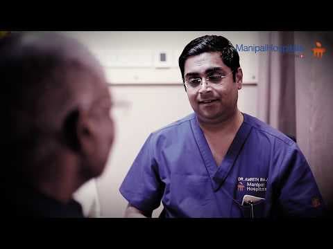
Robot Assisted Radical Prostatectomy
A miraculous Procedure, Robot Assisted Radical Prostatectomy done by Dr. Amrith Raj Rao, Consultant - Urology And Robotic Surgery...
With a legacy of several decades, Manipal Hospitals is one of India's leading multi-speciality healthcare networks. We deliver comprehensive outpatient, inpatient, emergency, and advanced surgical care tailored for patients of all age groups.

Not sure what you're looking for?
A gamma camera is a nuclear medicine imaging device that detects gamma rays emitted from a radioactive tracer inside the body, producing images that show how organs and tissues are functioning.
After a small dose of radioactive tracer is given, it travels to specific organs or tissues. The gamma camera detects the gamma rays released by the tracer and converts them into detailed functional images.
Yes. The radioactive tracer used is given in a very small, carefully controlled dose, and gamma camera radiology procedures are considered safe for most patients.
Gamma camera imaging is commonly used to evaluate bone conditions, thyroid function, kidney function, and heart perfusion, as well as to detect the spread of certain cancers.
Depending on the type of study, the scan can take anywhere from 20 minutes to a few hours, as time is needed for the tracer to circulate before imaging begins.